
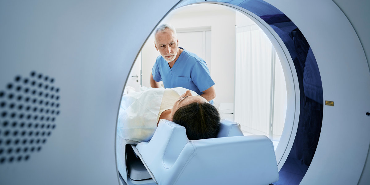
In today's rapidly advancing medical landscape, the role of imaging technology in early detection and prevention of diseases cannot be overstated. As healthcare professionals strive to improve patient outcomes and reduce the burden of chronic conditions, imaging technology has emerged as a key tool for diagnosing diseases in their earliest stages.
In an article published in Artificial Intelligence in Medicine in 2022, the author explained that according to the Journal of the American Medical Association, over 80% of visits to hospitals and healthcare systems involved a minimum of one imaging examination, resulting in an annual expenditure of approximately $65 billion on diagnostic imaging.
This powerful approach through different imaging modalities enables doctors to identify and treat various medical conditions before they become life-threatening, ultimately saving lives and improving overall quality of life.
Some areas which will be discussed in this article that are currently benefiting from imaging technology include cancer and neurodegenerative diseases.

Medical Imaging for Early Detection of Cancer
Medical imaging plays a significant role in early cancer diagnosis, particularly when it comes to screening both healthy and high-risk populations. The main goal of cancer screening is to detect the disease at an early stage, ideally before symptoms appear and metastasis occurs. This allows for more effective treatments, reduced morbidity, and improved survival rates. In some cases, early detection may even enable successful treatment through surgery alone.
However, according to an article published in the American Society of Clinical Oncology Educational Book in 2015, the authors considered it crucial to note that not all cancer types have well-established screening methods, and some tests may only provide marginal benefits when considering their potential harm. As such, there is an ongoing debate in the oncology community regarding the ideal timing and choice of imaging modalities to maximize benefits while minimizing risks.

The current review highlights three key aspects of cancer screening and early tumor surveillance: tumor biomarkers, imaging, and circulating tumor cells and DNA. Among these, radiographic imaging has been used for cancer screening for decades, with multiple clinical studies demonstrating its efficacy in specific cases.
Despite the ongoing debate about the optimal use of imaging, there is a consensus on the benefits of early cancer detection using imaging techniques for three main cancer types: breast cancer, colorectal cancer (CRC), and lung cancer.
Medical Imaging for High-Risk Populations - Breast and/or Ovarian Cancer
In the article mentioned above Early Detection of Cancer: Past, Present, and Future, the authors stated that medical imaging, particularly mammography, is essential for early detection of breast cancer. They shared a comprehensive review by Pace and Keating that, based on over 50 years of evidence, found that regular mammography screening reduces breast cancer mortality by 19% on average (15% for women in their 40s and 32% for women in their 60s).
However, the researchers also added that the risk of false-positive results is high, with over 60% of women undergoing annual mammograms in their 40s to 50s experiencing false positives, which leads to increased anxiety, biopsies, and medical costs.
Some organizations have proposed mammography guidelines, recommending that women aged 50 to 70 should receive a mammogram at least every two years, with some groups suggesting annual screenings from age 40. When discussing mammography with patients, it is important to emphasize that it is not a perfect test, but it does save lives. Mammography can overdiagnose and produce false positives, and informed decisions should consider family history, individual risk, preferences, and expert recommendations.
Women with hereditary breast and/or ovarian cancer (HBOC) syndrome, specifically those with BRCA1/BRCA2 mutations, have a lifetime breast cancer risk of 40% to 65%. The authors recommended screening for these women using mammography combined with breast MRI, as MRI offers better visualization of denser breast tissue often found in younger women.
Ironically, they also added that exposure to mammography before age 30 has been linked to an increased risk of breast cancer in women with BRCA1/BRCA2 mutations. Mammography plus breast MRI offers comparable survival benefits to prophylactic bilateral mastectomy at age 25 and prophylactic bilateral salpingo-oophorectomy at age 40.
Sensitivity, metastasis-free survival, and overall survival were higher in patients with familial breast cancer treated with MRI compared to mammography-based screening for invasive cancer.

Medical Imaging for Colorectal Cancer (CRC)
Colorectal cancer (CRC) is a significant health concern in the United States. The American Cancer Society published data with estimated numbers for the year 2023 regarding this disease. It is expected that by the end of the year there will be:
- 106,970 new cases of colon cancer
- 46,050 new cases of rectal cancer
- Over 52,550 CRC-related deaths
Early detection of colorectal cancer through medical imaging has been shown to improve clinical outcomes over the past four decades. Additionally, according to the authors of the article published in the American Society of Clinical Oncology Educational Book in 2015, the removal of adenomatous polyps, precursors to CRC, can dramatically reduce the risk of developing the disease.
In the same line, researchers added that regular colonoscopy screenings have been found to substantially reduce both CRC risk and mortality, with odds ratios and standardized mortality ratios ranging from 0.23 to 0.71.
The protective effects of colonoscopy screenings are more pronounced in the distal colon compared to the proximal colon. In the United States, gastroenterologists recommend CRC screening for the general population at five-year intervals starting at age 50. However, other guidelines suggest a 10-year interval may be just as effective for individuals at average risk.
Authors highlighted that the National Comprehensive Cancer Network (NCCN) recommends CRC screening at age 50 for those without a family history of the disease or personal history of adenoma, sessile serrated polyps (SSP), CRC, or inflammatory bowel disease. Screening methods include colonoscopy, stool-based guaiac/immunochemical testing, and flexible sigmoidoscopy. Rescreening recommendations vary based on the initial findings.
Despite widespread adoption of colonoscopy for CRC screening, the researchers from Early Detection of Cancer: Past, Present, and Future, noted that 6% of patients still developed interval tumors within 6 to 60 months of their colonoscopy.

For high-risk populations with hereditary CRC syndromes, early detection and polyp removal through rigorous screening programs are crucial. The NCCN provides specific recommendations based on the patient's mutation and associated CRC lifetime risk and earliest age of presentation. These guidelines vary depending on the genetic mutation and associated syndrome, with different recommendations for Lynch syndrome, familial adenomatous polyposis (FAP), attenuated FAP, MUTYH-associated polyposis (MAP), Peutz-Jeghers syndrome, and juvenile polyposis syndrome.
Medical Imaging for Lung Cancer
Lung cancer is the is the second most common cancer in both men and women in the United States. Data from the American Cancer Society estimates that by the end of the year 2023 there will be:
- Around 238,340 new cases of lung cancer (117,550 in men and 120,790 in women)
- Approximately 127,070 deaths from lung cancer (67,160 in men and 59,910 in women)
Advances in imaging technology have led to improved lung cancer screening and early detection through annual low-dose CT (LDCT) scans. Authors of the study published in the American Society of Clinical Oncology Educational Book in 2015, stated that the National Lung Screening Trial (NLST), the largest randomized clinical trial, demonstrated a 20% reduction in death for current or former smokers.
Additionally, other organizations such as the United States Preventive Services Task Force, American College of Chest Physicians/American Society of Clinical Oncology, American Association of Thoracic Surgeons, NCCN, American Cancer Society, and American Lung Association, have provided overlapping yet distinct recommendations for lung cancer screening.
Most of these organizations recommend annual LDCT for high-risk individuals, which includes patients aged 55 to 79 with a 30 pack-year smoking history or more, former smokers who quit within the past 15 years, or patients aged 50 to 79 with a 20 pack-year smoking history or more who have additional risk factors.
Cost-effectiveness analyses are being conducted for the national adoption of LDCT as the preferred method of screening for lung cancer in current or former smokers. In the same article, Early Detection of Cancer: Past, Present, and Future, authors mentioned one study that estimated healthcare expenditures to reach $1.3 to $2 billion with 50% to 75% screening uptake and $240,000 in additional costs to avoid one cancer death.

While LDCT may prevent over 8,000 annual deaths from lung cancer, careful cost-effectiveness analyses are crucial to understanding its true value. The Centers for Medicare & Medicaid Services (CMS) recently announced that Medicare will cover LDCT in current or previous smokers.
Medical Imaging for Neurodegenerative Diseases
Medical imaging has played a pivotal role in the diagnosis and monitoring of neurodegenerative diseases over the past twenty years. The inclusion of biomarkers in diagnostic procedures and clinical trials has revolutionized the understanding of these complex conditions. Neuroimaging biomarkers provide valuable insights into brain structure and function, allowing for better characterization and tracking of disease progression.
According to a study published in La Presse Médicale in 2022, there are different imaging modalities used for assessing neurodegenerative diseases, each with its unique advantages and applications. Magnetic Resonance Imaging (MRI) is primarily used to investigate brain structure, enabling healthcare professionals to visualize changes in brain tissue and identify potential abnormalities associated with the disease.
On the other hand, molecular imaging, functional MRI (fMRI), and electro- and magnetoencephalography (EEG and MEG) focus on examining brain function. These techniques can reveal information about neuronal activity, blood flow, and metabolic processes, which can help clinicians understand the functional consequences of neurodegenerative diseases and monitor treatment responses.
Medical Imaging for Alzheimer and Parkinson
Authors of Functional Imaging for Neurodegenerative Diseases highlighted that medical imaging techniques, such as MRI and PET scans, have become an essential part of managing neurodegenerative disorders like Alzheimer's and Parkinson's diseases. Previously, physicians relied solely on clinical approaches; however, the integration of neuroimaging biomarkers has allowed for better diagnosis and understanding of these conditions.
Researchers added that diagnosing dementia can be challenging, especially in its early stages or when symptoms overlap with other disorders. The increasing focus on biomarkers is helping to redefine dementia diagnosis based on biological factors rather than just clinical symptoms.

Neurodegeneration is believed to spread along neural pathways, leading to distinct patterns of cerebral pathologies. Multimodal neuroimaging studies have been crucial in identifying these patterns. However, it's important to consider neuroimaging biomarkers alongside clinical conditions and other biological biomarkers for a more accurate diagnosis, particularly in cases where early detection is difficult, or disease progression is subtle.
According to the same study mentioned above, advanced MRI techniques are valuable in characterizing specific neurodegenerative conditions and monitoring disease progression. These techniques range from simple visual inspection of MRI scans to more complex methods like volume measurements, diffusion tensor MRI, and functional MRI. Morphological MRI sequences can identify structural changes and patterns of atrophy in neurodegenerative diseases, while diffusion tensor imaging (DTI) and resting state functional MRI can analyze functional connections between brain areas.
PET scans are widely used to assess regional glucose utilization patterns, which can help differentiate between various neurodegenerative subtypes. Recent advancements in radiopharmaceuticals and PET tracers targeting misfolded proteins enable quantitative and anatomical analysis of protein presence and change over time. This ability to measure amyloid burden offers new diagnostic and treatment monitoring possibilities. Additionally, the development of Tau and α-synuclein specific tracers enhances the pathophysiological understanding of these diseases.
The authors of the study added that both PET and MRI imaging offer surrogate biomarkers for neurodegenerative disorders. MRI is non-invasive, cost-effective, and repeatable, while PET scans can provide more specific information about misfolded proteins. Despite the numerous potential biomarkers under investigation, there remains a gap between these emerging techniques and the limited number of validated biomarkers used in patient diagnosis and covered by health insurance.
Tau Protein Detection and Tracking
According to another study published in the European Journal of Nuclear Medicine and Molecular Imaging in 2015, a key focus of medical imaging is the detection and tracking of tau protein, which when aggregated, becomes a significant pathological factor in Alzheimer and Parkinson.
Tau is a protein that helps stabilize neurons and aid in the transport of axonal nutrients. However, when it becomes hyperphosphorylated, it aggregates and leads to conditions known as tauopathies, which include Alzheimer's disease (AD) and Parkinson's disease (PD). In AD, tau tangles are closely linked with neuronal dysfunction and cell death, more so than β-amyloid plaques. Indeed, tau deposition is more closely associated with symptom onset due to neuronal dysfunction, with tau levels at autopsy correlating well with pre-morbid cognitive status.

In the same study, the authors explained that in the past, information about tau deposition was only available through invasive techniques such as brain biopsy or autopsy. However, recent advances in selective tau tracer development for positron emission tomography imaging have allowed for the in-vivo exploration of the presence and extent of tau pathology in patients suspected of having tauopathies.
These advances in tau PET imaging offer support in the early differential diagnosis of neurodegenerative disorders by revealing whether a characteristic pattern of aggregated tau is present. It also provides a potential biomarker of disease progression, which can be crucial for informing treatment plans and understanding the likely course of the disease.
The National Institute of Ageing–Alzheimer’s Association (NIA–AA) Working Group Guidelines highlight the concept of AD as a spectrum or continuum of disease, with pathophysiological changes occurring long before the onset of cognitive symptoms and ultimate dementia. During the asymptomatic prodrome, imaging biomarkers can potentially be used to stage disease and follow its progression.
The authors of Tau Imaging in Neurodegenerative Diseases concluded that while β-amyloid imaging plays a key role in the evaluation of dementia, the closer correlation of tau with cognitive impairment and neuronal dysfunction makes it more suitable as a biomarker of disease progression.
The development of novel tau tracers and ongoing clinical studies are anticipated to not only extend our knowledge of dementia but also to diagnose dementia accurately and evaluate multi-targeted therapy more efficiently. Thus, medical imaging in the context of Alzheimer's and Parkinson's disease continues to evolve, promising significant contributions to our understanding and management of these debilitating conditions.

These discussed advancements in imaging technology have revolutionized the medical field, providing invaluable tools for early detection and prevention of diseases like cancer, Alzheimer's, and Parkinson's. Techniques such as PET scans, MRI, and advanced tau tracers offer insights into the human body that were once impossible to obtain without invasive procedures.
These technologies are not only helping to identify diseases at their earliest stages when they are most treatable, but they're also providing detailed information about disease progression and response to treatment. This is particularly crucial in neurodegenerative diseases, where early intervention can significantly alter the disease trajectory and improve a patient's quality of life.
The role of biomarkers in disease detection and management is becoming increasingly critical. With more research and development, we will continue to see even more refined imaging techniques and biomarkers, leading to better, more personalized care for patients. As we move forward, it's clear that imaging technology will continue to be an essential weapon in our healthcare arsenal, helping us to detect, understand, and ultimately conquer these challenging diseases.
Sources
Chollet, François and Payoux, Pierre “Functional Imaging for Neurodegenerative Diseases”La Presse Médicale Vol. 51, Iss. 2 June 2022
Connecting imaging and care delivery to improve outcomes, 2022
https://ai-med.io/analysis/technology/connecting-imaging-and-care-delivery-to-improve-outcomes/
Dani, M. et al “Tau imaging in neurodegenerative diseases” European Journal of Nuclear Medicine and Molecular Imaging Vol. 43 1139–1150 2016
Key Statistics for Colorectal Cancer, 2023
https://www.cancer.org/cancer/types/colon-rectal-cancer/about/key-statistics.html
Key Statistics for Lung Cancer, 2023
https://www.cancer.org/cancer/types/lung-cancer/about/key-statistics.html
Schiffman, Joshua et al. “Early Detection of Cancer: Past, Present, and Future.” American Society of Clinical Oncology Educational Book, Vol. 35 57-65 2015